Many clinicians consider fluoroscopy an antiquated tool, but CT scans can cause harmful side effects. MRIs are often tricky to schedule quickly. Ultrasounds also have their limitations. Doctors need guidance on which test to recommend for diagnosing specific digestive conditions.
But does it matter which imaging exam to prescribe?
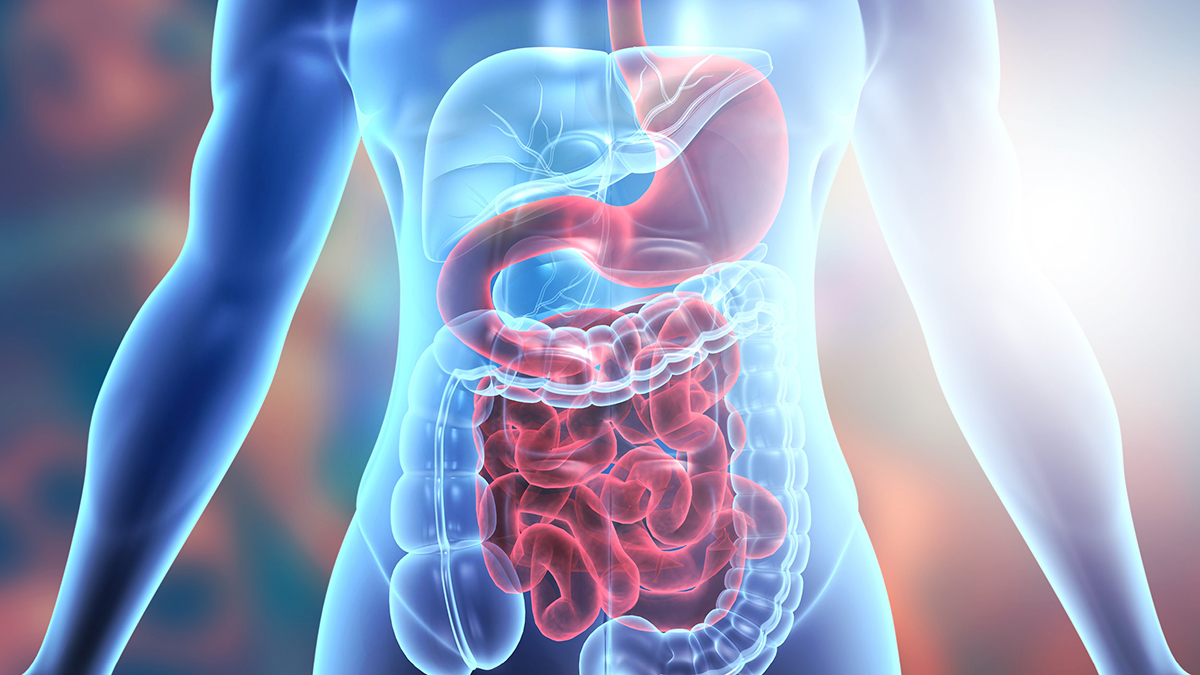
Inappropriate imaging can lead to incorrect diagnoses and therapies. Additionally, it can produce harmful side effects. With the expansion and evolution of radiology services, deciding which exam to prescribe can be challenging. Physicians often question which exam would be best for diagnosing digestive issues. This article provides a brief comparison of four imaging tools that aid in diagnosing digestive issues.
Ultrasounds are typically the first exam physicians use to diagnose digestive issues. This test is frequently sufficient for a complete diagnosis. It boasts portability and accessibility. Studies show that ultrasounds provide 40% more information than clinical assessment alone. Based on the information obtained, physicians modify management strategies 20% of the time. Research indicates that ultrasounds for acute abdominal pain reduce emergent CT scans by 50%. With the advancements of contrast ultrasounds, diagnostic capabilities have significantly improved.
So, when should clinicians utilize ultrasounds for diagnosing digestive issues?
Though ultrasound imaging provides many advantages, it does have some limitations. Unlike MRIs and CT scans, ultrasounds do not have cross-sectional capabilities. Sound waves don’t travel well through gas or bone. This factor limits the visualization of gas-filled organs or tissue surrounded by bone. Further, ultrasound imaging does not detect objects or organs deep in the body.
The GI series is a group of X-ray exams that use fluoroscopy to visualize organs along the GI tract. Physicians may recommend one or all the tests of the GI series, depending on the part of the tract affected. Fluoroscopy uses continuous X-ray beams to make a real-time movie. This capability delivers in-action visualization of a specific body part.
Fluoroscopic exams for diagnosing digestive issues are the following:
For maximum clarity, fluoroscopy requires contrast. Physicians typically use barium as the contrast agent in an upper GI series. Fluoroscopy is a viable imaging option for the following abdominal conditions:
Like ultrasound imaging, fluoroscopy also has some limitations. It uses radiation. However, compared to a CT scan, the dosage is significantly less. It is not a high-resolution resource and does not produce cross-sectional views. Fluoroscopy currently delivers a 2-D view. However, innovators are improving this imaging modality to make 3-D fluoroscopy more accessible.
Computed tomography provides superior morphological resolution and a cross-sectional view. Additionally, this tool allows 3-D imaging of multiple structures with one exam. For example, physicians can visualize pelvic organs, bones, lungs and abdominal viscera with a single test.
CT scans are essential for evaluating the abdomen during severe emergencies. This tool also helps in staging patients with cancer. Due to its many advantages, physicians frequently rely on CT for diagnostic help. However, this exam should not be performed haphazardly. CT scans use ionizing radiation; therefore, overuse can produce harmful, long-term side effects. Physicians should utilize ultrasound or fluoroscopic imaging for low-risk conditions.
Computed tomography provides excellent imaging for the following GI tract conditions:
CT imaging is a valuable tool, but it also has limitations. Its ionizing radiation prevents it from being the go-to resource for all digestive conditions. Additionally, it requires patients to be hemodynamically stable and able to lie still.
MRIs play a significant role in the evaluation of GI tract conditions. They deliver excellent soft-tissue visualization and a 3-D, cross-sectional view. This view displays the total thickness of the gastric wall. It helps physicians evaluate the deep pelvis ileal loops without superimposition. It provides visualization of the mesentery and pericentric fat. Additionally, this exam can assess solid organs and supply an overview of the abdomen and pelvic region. Magnets, radio frequencies and a computer deliver detailed images without ionizing radiation. It is helpful for specific conditions such as:
Accessibility is a significant limitation for MRI use. Physicians often complain that MRI imaging is challenging to utilize. Further, extensive exam time prevents physicians from using MRIs during abdominal emergencies.
With abundant imaging options, physicians need support. What once was the go-to resource for imaging information may now be an antiquated option. Inappropriate imaging can lead to misdiagnosis and incorrect intervention. As an attentive clinician, you want to be confident you’re choosing the proper exam for the presenting condition.
We understand that advancements are ever-evolving. Change and innovation produce unique challenges. We are here to help you maximize resources, improve efficiency and advance efficacy. To learn more about our imaging resources, click the “Refer” button.
“Indications for abdominal imaging: When and what to choose?.” NIH: National Library of Medicine, 2020, Indications for abdominal imaging: When and what to choose? – PMC.
“X-Ray Studies of the Digestive Tract.” Merck Manual Consumer Version, 2023, X-Ray Studies of the Digestive Tract – Digestive Disorders – Merck Manual Consumer Version.
“Gastrointestinal imaging-practical magnetic resonance imaging approach.” NIH: National Library of Medicine, 2014, Gastrointestinal imaging-practical magnetic resonance imaging approach – PMC.
Subscribe to our MEDforum e-newsletter for the latest guidelines and information from Up Health.