Skip to site content

Over the past couple of decades, the clinical use of fluoroscopy has declined significantly. So, is it still an essential diagnostic modality? Many emerging physicians say, “No,” discounting it as a viable resource.
However, the National Institute of Health (NIH) states “Fluoroscopy remains a valuable modality, even in the age of high-resolution cross-sectional imaging modalities such as computed tomography (CT), magnetic resonance imaging (MRI), and ultrasound.” The NIH emphasizes its importance. That’s why they call for a better understanding of its place in diagnostic radiology. Keep reading to learn why and how to utilize this essential imaging tool.
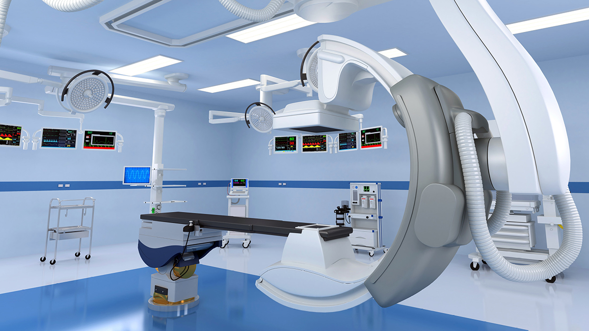
Fluoroscopy has come a long way since its invention in 1896 by Thomas Edison. What once was a dangerous procedure is now a safe diagnostic imaging solution. The fluoroscopy delivers continuous X-ray imaging. The X-ray beam passes through the body, providing a real-time movie.
Fluoroscopy helps physicians visualize the organ function of a specific body part. This functional exam aids with diagnosing and treatment planning. Over the past few decades, the clinical use of fluoroscopy has declined for several reasons.
The abundance of imaging options can make the prescribing decision challenging. With the development of high-resolution modalities, imaging options have expanded over the past century.
Computed tomography (CT), magnetic resonance imaging (MRI) and ultrasounds deliver many diagnostic advantages. Physicians now utilize focused ultrasound, MRI enterography, and abdominal CT to see pathologies previously visualized with fluoroscopy. However, the NIH claims that fluoroscopy remains an essential diagnostic tool, delivering unique capabilities.
The abundance of imaging options is not the only factor contributing to the decline in fluoroscopy use. Medicare price structure affects differential reimbursement per procedure performed. Financial reimbursement rates are less for fluoroscopy than for CT or MRI exams. This factor plays a significant role in decision-making for diagnostic radiology.
Emerging medical professionals often consider fluoroscopy an antiquated form of imaging. They favor CT scans and MRIs, discounting fluoroscopy as a valuable imaging option. This is in part due to the limited training related to fluoroscopy. Many lack confidence in when or how to utilize it.
CT scans and MRIs usually need only a technician to perform the exam. However, fluoroscopy requires a trained radiologist to perform the procedure and interpret the results. This factor makes fluoroscopy more challenging to access.
Physicians are constantly evaluating the benefits and risks of treatment options. Radiology services are vital for diagnosing illnesses and strategizing interventions. So, what are the benefits of fluoroscopy?
Fluoroscopy provides quick real-time evaluation with true temporal resolution. Physicians can noninvasively visualize the movement and function of many organs. This feature helps physicians deliver prompt assessments and therapies, improving patient experience and outcomes.
Fluoroscopy also allows for patient repositioning during the exam. This feature is not typically available with traditional CTs and MRIs. Physicians can adjust the device for optimal visualization. This capability delivers a precise and functional radiology tool.
Fluoroscopy provides less ionizing radiation exposure compared to a CT scan. The average dose of a typical fluoroscopy is 10-50 mGy/min with an exam time of a minute or less. A CT of the pelvis region delivers approximately 12mSv of radiation, and a multiphase CT of the pelvis region is up to 24 mSv. Fluoroscopy is a viable imaging option for those concerned about radiation exposure.
These benefits are why fluoroscopy adds value to the imaging team. It is useful for routine diagnostic services and in combination with other imaging resources.
Fluoroscopy should be routinely used to help visualize structural and functional anatomy. It is the preferred imaging for the initial workup of the following:
Due to low radiation dosage, fluoroscopy is the imaging modality of choice for pediatric patients. When evaluating tubal patency, fluoroscopy delivers high percentages related to sensitivity (92.1%) and specificity (85.7%).
Fluoroscopy is also a valuable resource when used synergistically with other imaging tools. Many situations call for a repeat CT scan. However, the elevated radiation levels make this inadvisable. Physicians can turn to fluoroscopy for a noninvasive, low-dose imaging option. When initial imaging is insufficient or the cross-sectional view is inconclusive, physicians should consider fluoroscopy.
Fluoroscopy remains an essential imaging modality, even in an age of high-resolution resources. Patient repositioning and reduced radiation exposure are beneficial to patients and providers. Additionally, its real-time visualization provides fast diagnostic results.
We utilize various imaging resources to decrease harm and improve patient care. Are you looking to partner with a facility that excels in its radiology services? We are here to help. Click the “Refer” button to get started.
“Fluoroscopy: An essential diagnostic modality in the age of high-resolution cross-sectional imaging.” NIH: National Institute of Health, 2020, Fluoroscopy: An essential diagnostic modality in the age of high-resolution cross-sectional imaging – PMC.
“Fluoroscopy.” U.S. Food and Drug Administration, 2023, Fluoroscopy | FDA.
“Radiation in Healthcare: Fluoroscopy.” Centers for Disease Control and Prevention (CDC), 2021, Radiation in Healthcare: Fluoroscopy | Radiation | NCEH | CDC.
Subscribe to our MEDforum e-newsletter for the latest guidelines and information from Up Health.